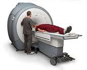
Recommendations presented for MRI use in multiple myeloma

(每日健康)建议magneti的使用c resonance imaging (MRI) in multiple myeloma are presented in a consensus statement published online Jan. 20 in theJournal of Clinical Oncology.
Meletios A. Dimopoulos, M.D., from the National and Kapodistrian University of Athens School of Medicine in Greece, and colleagues used data published through March 2014 to develop recommendations for the value of MRI inmultiple myeloma.
The researchers note that compared with other radiographic methods, MRI has high sensitivity for the early detection of marrow infiltration by myeloma cells. MRI detects bone involvements earlier than the myeloma-related bone destruction and without radiation exposure. MRI is the gold standard for axial skeleton imaging, evaluation of painful lesions, and differentiation of benign and malignant osteoporotic vertebral fractures. MRI can detect spinal cord or nerve compression and presence of soft tissue masses and is recommended for solitary bone plasmacytoma workup. All patients should undergo whole-body MRI or spine and pelvic MRI for smoldering or asymptomatic myeloma. MRI can provide prognostic information at diagnosis of symptomatic patients and after treatment.
"MRI describes the pattern of myelomatous infiltration of the bone marrow and is the procedure of choice for the evaluation of painful lesions in patients with myeloma for the detection of spinal cord compression and differentiation of malignant from nonmalignantvertebral fractures," the authors write.
Several authors disclosed financial ties to the pharmaceutical industry.
More information:Abstract
Full Text (subscription or payment may be required)
Editorial
Copyright © 2015HealthDay. All rights reserved.


