New brainstem model reveals how brains control breathing

Scientists from Karolinska Institutet, Sweden, have discovered how the brain controls our breathing in response to changing oxygen and carbon dioxide levels in the blood.
The control ofbreathingis essential for life. Without an adequate response to increased carbon dioxide levels, people can suffer from breathing disturbances, sickness, and panic. In worst-case scenarios, it can lead to premature death, as in sudden infant death syndrome.
There has been some debate over how the brain controls breathing. Now, a new study in mice, to be published in the journaleLife, shows that when exposed to decreased oxygen or increased carbon dioxide levels, the brain releases a small molecule called Prostaglandin E2 (PGE2) to help protect itself and regulate breathing.
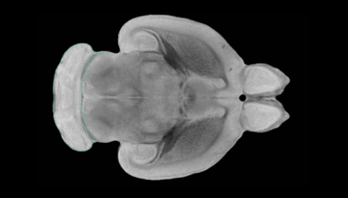
To discover this mechanism, the researchers grew a section of a mouse's brainstem (the central trunk of the brain) in a type of dish. The slice contained an arrangement of nerve and supporting cells that allowed it to 'breathe' for three weeks. During this time, the team monitored the cells and their behaviour in response to changes in the environment.
"Our novel brainstem culture first revealed that cells responsible for breathing operate in a small-world network. Groups of these cells work very closely with each other, with each group interconnected by a few additional cells that appear to work as hubs. This networking activity and the rhythmic respiratory motor output it generated were preserved for the full three weeks, suggesting that our brainstem can be used for long-term studies of respiratory neural network activity," explains David Forsberg, PhD student and first author of the study.
"Secondly, we saw that exposure to different substances made the brainstem breathe faster or slower. Perhaps most interesting was its response to carbon dioxide, which triggered a release of PGE2. Here, PGE2 acted as a signaling molecule that increased breathing activity in the carbon dioxide-sensitive brainstem region, leading to slower and deeper breaths, or 'sighs'."
These new insights have important implications for babies, who experience significantly reduced levels of oxygen during birth. At this stage, PGE2 protects the brain and prepares the brainstem to generate deep sigh-like breath intakes, resulting in the first breaths of air following birth.
The study also reveals a novel pathway linking the inflammatory and respiratory systems. PGE2 is released during inflammation and fever, which can dysregulatebreathing patternsand interfere with normal responses to carbon dioxide. This can in turn cause disturbed and even dangerous halts in breathing.
"Our findings go some way to explaining how and why our breathing responses to imbalanced oxygen andcarbon dioxidelevels are impaired during infectious episodes. It also helps further our understanding of why infection can inhibit breathing so severely in new-born babies," says Eric Herlenius, Professor at Karolinska Institutet's Department of Women's and Children's Health, and senior author of the paper.
"We now want to find out how breaths form and develop during episodes of inflammation. This could be useful for researching potential new ways to save babies' lives when they are unable to catch their breaths."
More information:David Forsberg et al, CO-evoked release of PGE2 modulates sighs and inspiration as demonstrated in brainstem organotypic culture,eLife(2016).DOI: 10.7554/eLife.14170


