特殊的转录因子及其靶基因是罕见白血病的治疗途径

间变大细胞淋巴瘤(ALCL)是一种主要发生在儿童和年轻人的白血病。MedUni Wien的一个国际研究团队现在已经能够证明转录因子BATF3及其靶基因在肿瘤细胞的生长中发挥关键作用。这项研究的发现,目前发表在自然通讯可以作为开发新疗法的一种方法。
恶性淋巴瘤是淋巴癌最常见的形式,如果淋巴细胞分裂不受控制。区分霍奇金淋巴瘤(HL)和非霍奇金淋巴瘤,其中还包括不常见的间变性大细胞淋巴瘤(ALCL),一种恶性t细胞淋巴瘤,特别影响儿童和年轻人。化疗是标准治疗方法,但经常复发。
BATF3是ALCL信号传递的关键转录因子
维也纳医学大学的一组研究人员,包括临床病理研究所的Olaf Merkel和Lukas Kenner(也在维也纳兽医大学),以及机构合作者,现在已经研究了转录因子BATF3在ALCL中的作用。它在ALCL肿瘤细胞中的消除对细胞生长有巨大的影响,这表明它是该疾病的重要蛋白质。研究人员对BATF3的兴趣是受到这些发现和BATF3深刻的疾病特异性表达的启发。
发现超增强子区域
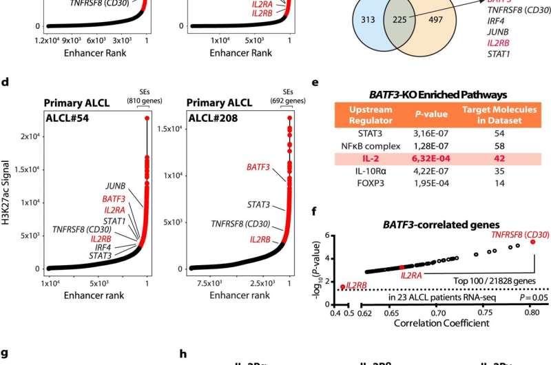
其中,Stephan Mathas周围的小组在早期的合作项目中已经证明了AP-1转录因子家族,包括JUNB、cJUN和BATF3在ALCL中严重表达。鉴于BATF3对疾病的重要性及其高表达,研究人员假设它可能位于基因组的所谓超级增强子区域。超级增强子是基因组中对基因调控和细胞识别具有重要意义的区域。与波士顿Tom Look实验室一起进行的h3k27组蛋白乙酰化全基因组分析证实,BATF3确实位于所有分析细胞系的超级增强子区域,重要的是在原发性ALCL患者样本中也存在。
此外,研究人员对BATF3芯片BATF3进行了全基因组结合测试,并确定BATF3与自己的启动子结合,从而产生正反馈环。Olaf Merkel解释说:“虽然我们观察到基因的表达被batf3敲除改变,但IL-2R系统的基因是最明显的,这促使我们在表达和功能方面仔细检查三聚体IL-2受体的成员。”
研究人员确定,ALCL中IL-2R复合体的所有三个亚单位都被严重激活,并且IL-2R α和- β是BATF3的直接靶标。“IL-2是t细胞激活后释放的最重要的白介素,”Merkel解释说,“我们能够证明IL-2能够促进ALCL肿瘤细胞的生长。在超过80%的ALCL患者中,所有三种IL-2受体亚基的高激活支持了IL-2对ALCL生长具有重要功能的观点,这与功能分析一起表明了IL-2信号在ALCL中的高度重要性。”另一个已知与IL-2受体共享两个亚基的密切相关的细胞因子是IL-15。研究人员也能在ALCL细胞上显示出促进生长的作用。
鉴于可能的治疗方法,研究人员研究了针对IL-2受体亚单位的武装抗体的效果,该抗体与针对ALCL细胞的细胞毒素偶联。即使单次给药这种抗体细胞毒素缀合物也能大量减少ALCL的肿瘤生长细胞在动物模型中,这可能是人类临床试验的基础。目前出版的研究结果有助于理解的发展和增长间变性大细胞淋巴瘤并有助于有效疗法的发展。
进一步探索


