一组老鼠表现出强迫性吸食可卡因的行为。(A)鉴定强迫性可卡因使用大鼠的实验时间表。(B)基于惩罚阶段2和3 (P2和P3)参数的t分布随机邻居嵌入(t-SNE)投影和包括抗性(集群1)和敏感(集群2)大鼠的集群t-SNE三维表示的分层聚类。行为表现包括主动刺鼻(A),不主动刺鼻(IA),无效刺鼻和可卡因注射。(C)敏感大鼠和抵抗大鼠的比例(左),以及基线和第三次惩罚阶段的可卡因注射(右)。与耐药大鼠相比,敏感大鼠输注较少。双向方差分析(ANOVA)显示交互作用有统计学意义,F1,89 = 80.76, P < 0.001,事后分析,P3敏感组与耐药组***P < 0.001, P3敏感组与耐药组与基线组###P < 0.001;N = 60和31。(D和E)从获取过程中获得的积极的、不活跃的和可卡因注射的鼻塞数量。敏感鼠和抵抗鼠之间没有差异; two-way ANOVA showed nonsignificant main effects on groups, F1,89 = 0.53, P = 0.4671 for active nosepokes, F1,89 = 0.03, P = 0.8639 for inactive nosepokes, and F1,89 = 0.14, P = 0.7086 for infusions; n = 60 and 31, respectively. NS, not significant. Data are presented as mean values ± SEMs. Credit:科学的进步(2022)。DOI: 10.1126 / sciadv.abq5745
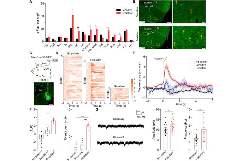
抗休克大鼠aIC神经元过度活跃。(A)第三次惩罚测试后90分钟大脑区域c-Fos的映射。OFC的未配对t检验,t11 = 2.24, *P = 0.046;t10 = 4.21, **P = 0.005;NAc岩心t12 = 2.50, *P = 0.039;NAc壳t12 = 2.53, *P = 0.027;t12 = 4.13, **P = 0.004为DLS;t12 = 2.71, *P = 0.03;N = 5 ~ 6, 7 ~ 8。(B) aIC c-Fos免疫荧光染色的代表性图像。 Scale bars, 500 μm (left) and 50 μm (right). (C) Schematic of calcium photometry. Scale bar, 1 mm. (D and E) Heatmap and plot of z-scored aIC Ca2+ signals in no punish, sensitive, and resistant group; n = 5 for each group. (F) AUC of z-scored aIC Ca2+ signals in 0 to 2 s. One-way ANOVA, F2,12 = 17.58, P < 0.001, post hoc analysis, *P = 0.028 for sensitive versus resistant, ***P < 0.001 for no punish versus resistant. Number of events per minute of aIC neurons. One-way ANOVA, F2,12 = 36.00, P < 0.001, post hoc analysis, ***P < 0.001 for sensitive versus resistant and no punish versus resistant; n = 5 for each group. (G) Amplitude and frequency of sEPSCs of aIC neurons. Unpaired t test, t32 = 2.12, *P = 0.042 and t32 = 3.60, **P = 0.001, respectively; 13 neurons from 3 sensitive rats and 21 neurons from 5 resistant rats. Cg1, cingulate cortex area 1; Cg2, cingulate cortex area 2; PrL, prelimbic cortex; IL, infralimbic cortex; NAc core/shell, nucleus accumbens core/shell; MS, medial septum; DLS, dorsolateral striatum; BLA, basolateral amygdala; CeA, central amygdala; PVT, paraventricular thalamus; LHb, lateral habenula. Data are presented as mean values ± SEMs. Credit:科学的进步, DOI: 10.1126/sciadv.abq5745
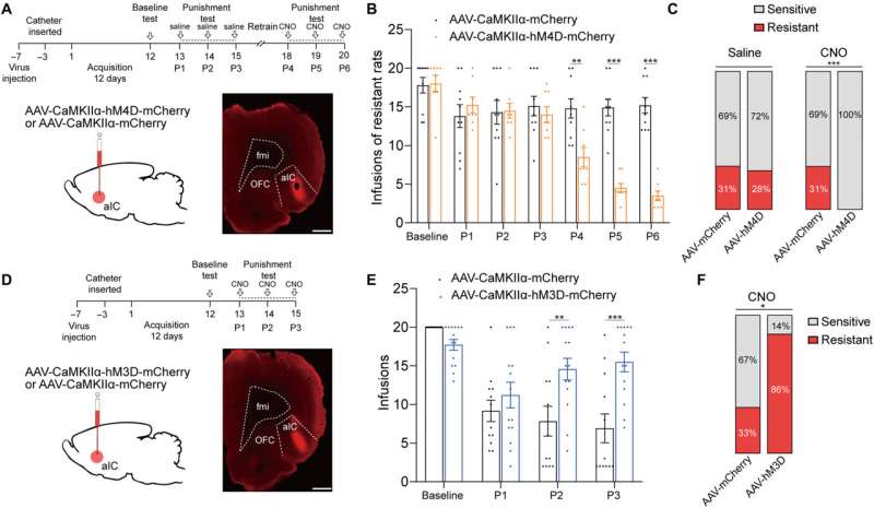
aIC神经元的激活对于强迫性可卡因使用是必要的,也是足够的。(A)病毒输注的实验时间表和示意图,以分析抑制aIC后耐药大鼠的强迫性可卡因使用行为。(B) AAV-mCherry组和AAV-hM4D组的可卡因注射液。双因素方差分析,F1,18 = 22.51, P < 0.001,事后分析,P4, P5, P6 ***P < 0.001;每组N = 10。(C) aIC抑制前后AAV-mCherry组和AAV-hM4D组大鼠敏感和耐药比例;Fisher精确检验,***P < 0.001。(D)病毒输注的实验时间表和示意图,分析aIC激活后的强迫性可卡因使用行为。鳞片条,1mm。(E)可卡因输注从AAV-mCherry和AAV-hM3D组获得。双向方差分析,F1,25 = 15.22, P < 0.001,事后分析,P2和P3的***P < 0.001; n = 14 and 13, respectively. (F) Ratio of sensitive and resistant rats in AAV-mCherry and AAV-hM3D group after aIC activation; Fisher's exact test, **P = 0.004. Data are presented as mean values ± SEMs. Credit:科学的进步, DOI: 10.1126/sciadv.abq5745
aIC谷氨酸神经元双向调节强迫性可卡因使用。(A)病毒输注的实验时间表和示意图,以分析耐药大鼠aIC谷氨酸神经元抑制后的强迫性可卡因使用行为。(B) AAV-mCherry组和AAV-hM4D组的可卡因注射液。双因素方差分析,F1,16 = 9.89, P = 0.006,事后分析,P4 **P = 0.001, P5、P6 ***P < 0.001;N = 10和8。(C) AAV-mCherry组和AAV-hM4D组大鼠抑制aIC谷氨酸能神经元前后敏感和抵抗的比例;Fisher精确检验,***P < 0.001。(D)病毒输注的实验时间线和示意图,以分析aIC谷氨酸神经元激活后的强迫性可卡因使用行为。鳞片条,1mm。(E) AAV-mCherry组和AAV-hM3D组的可卡因注射液。双因素方差分析,F1,24 = 6.42, P = 0.018,事后分析,P2 **P = 0.004, P3 ***P < 0.001; n = 12 and 14, respectively. (F) Ratio of sensitive and resistant rats in AAV-mCherry and AAV-hM3D group after activation of aIC glutamatergic neurons; Fisher's exact test, *P = 0.014. Data are presented as mean values ± SEMs. Credit:科学的进步, DOI: 10.1126/sciadv.abq5745
在耐药大鼠中,从OFC到aIC的谷氨酸投射增强。(A)接受OFC输入的aIC神经元的全细胞膜片钳记录的实验时间轴和示意图。比例尺,左为1mm,右为100 μm。(B) OFC-aIC神经元记录的sEPSCs振幅和频率。无配对t检验,振幅t35 = 2.42, *P = 0.021;t35 = 10.71,频率***P < 0.001, 21个神经元来自4只敏感大鼠,16个神经元来自4只抗性大鼠。(C)从接受OFC输入的aIC神经元获得的响应增加阶跃电流的ap数量。双因素方差分析,f1187 = 297.4, P < 0.001,事后分析,200 ~ 600 pA *P < 0.05, 4只敏感大鼠18个神经元,3只抗性大鼠19个神经元。(D)全细胞膜片钳记录aIC神经元对470纳米激光反应的实验时间线和示意图(2ms,蓝色条)。(E和F)与不惩罚和敏感大鼠相比,抗性大鼠aIC神经元PPR显著降低。 One-way ANOVA, F2,45 = 4.66, P = 0.0145, post hoc analysis, *P = 0.0375 for sensitive versus resistant, and *P = 0.0199 for no punish versus resistant. (G and H) The A/N ratio of aIC neurons was significantly increased in resistant rats. One-way ANOVA, F2,45 = 54.36, P < 0.001, post hoc analysis, ***P < 0.001 for sensitive versus resistant and no punish versus resistant; 16 neurons from three no punish rats, 18 neurons from three sensitive rats, and 14 neurons from three resistant rats. Data are presented as mean values ± SEMs. Credit:科学的进步, DOI: 10.1126/sciadv.abq5745
 Experimental timeline for identifying rats with compulsive cocaine use. (B) Hierarchical clustering based on t-distributed stochastic neighbor embedding (t-SNE) projection of parameters in punishment sessions 2 and 3 (P2 and P3) and t-SNE three-dimensional representation of clusters including resistant (cluster 1) and sensitive (cluster 2) rats. Behavioral performance included active nosepokes (A), inactive nosepokes (IA), futile nosepokes, and cocaine infusions. (C) Ratio of sensitive and resistant rats (left), and cocaine infusions obtained from baseline and the third punishment session (right). Sensitive rats had fewer infusions compared with resistant rats. Two-way analysis of variance (ANOVA) revealed a statistically significant interaction effect, F1,89 = 80.76, P Science Advances (2022). DOI: 10.1126/sciadv.abq5745")