RMS的scRNAseq识别肌肉发育程序的异质性。(一)实验工作流程。(B)回归计数RNA数量、线粒体基因百分比和运行批处理效应后48,859个RMS细胞的UMAP图。细胞根据相应的样品来源进行颜色编码。(C)整合后48859个RMS细胞的UMAP图。所示为鲁汶聚类所识别的种群。(D)显示aRMS和eRMS样本中不同鲁汶聚类中谱系特异性标记基因表达的点图。(E)在RMS中确定的种群的骨骼肌生成模型。UMAP图以描述肌源性谱系进展的标记物的表达为基础进行着色。(F)整合后的RMS细胞UMAP图,并根据样本来源进行颜色编码。 (G) Relative proportion of Louvain clusters. Data are represented as means ± SEM; ordinary two-way analysis of variance (ANOVA) with uncorrected Fisher's least significant difference (LSD). *P < 0.05; **P < 0.01; ****P ≤ 0.0001. (H and I) Comparison of intratumoral heterogeneity between preclinical models of aRMS. O-PDXs and patient tumor data are derived from reference (36). UMAP plots of individual models (H) and of the integrated datasets (I) are shown. (J) Relative proportion of Louvain clusters across different aRMS preclinical models and patient tumors. Data are represented as means ± SEM of n = 6 patient tumors, n = 5 O-PDXs, n = 5 primary cultures, and n = 3 cell lines. Credit:科学的进步(2023)。DOI: 10.1126 / sciadv.ade9238
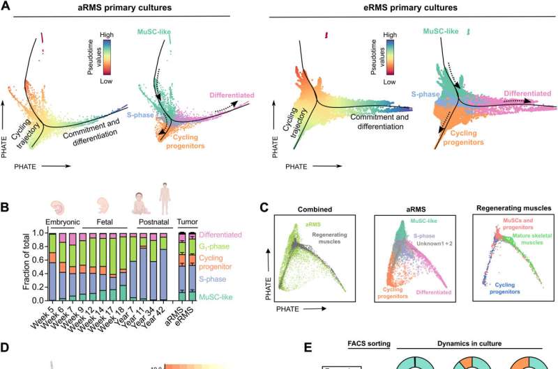
RMS原代培养重现了分枝性肌源性轨迹。(A) aRMS(左)或eRMS(右)原代培养的PHATE降维图(t = 30, knn = 20)。黑线表示使用Slingshot计算的伪时间轨迹(起始群集:类musc);虚线箭头表示轨迹方向。细胞根据Slingshot(左)计算的伪时间值或确定的Louvain集群(右)进行着色。(B)综合RMS/人类发育骨骼肌(42)数据集在发育时间点或RMS子类型上的聚类分布。(C)综合aRMS原代培养/小鼠再生骨骼肌(32)数据集的PHATE降维图(t = 30, knn = 20)。aRMS细胞在鲁汶簇的基础上进行颜色编码;肌肉细胞在原始出版物中确定的集群的基础上着色。(D)基于CD44(绿色)或MYOG(橙色)表达(左图)着色的aRMS原代培养物PHATE降维图(t = 30, knn = 20)。 The two markers are mutually exclusive (right plot). (E) Flow cytometry analysis of sorted CD44+ and CD44− subpopulations in aRMS-3 cells. Unsorted reference is also shown. Data are represented as means of n ≥ 2 biological replicates. (F) Relative proportion of Louvain clusters across aRMS-1 and aRMS-3 cells before sorting. The percentage of differentiated cells in the CD44− subpopulation is shown. (G) Immunofluorescence analysis of MyHC expression 7 days after sorting of CD44+ and CD44− subpopulations in aRMS-1 cells. The percentage of MyHC+ cells is indicated on the top right of each panel. (H) Proposed model of aRMS hierarchical structure compared to developing or regenerating MuSCs. Credit:科学的进步(2023)。DOI: 10.1126 / sciadv.ade9238
Science Advances (2023). DOI: 10.1126/sciadv.ade9238">
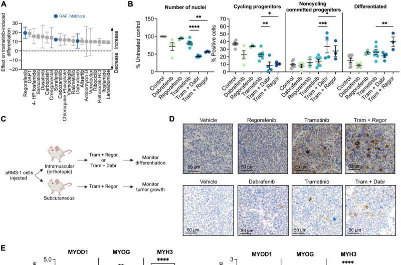
垂直抑制RAF-MEK-ERK级联增强曲美替尼诱导的分化并抑制aRMS肿瘤生长。(A)曲美替尼增强药物对曲美替尼诱导分化的影响排名前20位。0分代表曲美替尼单独的基线评分;阳性评分的药物增强曲美替尼诱导的分化。N = 2个生物重复。(B) aRMS-1细胞暴露于车辆对照、10 μM达格拉非尼(Dabr)、1 μM瑞格拉非尼(Regor)、10 nM曲美替尼(Tram)或指示组合72小时的免疫荧光定量分析;带未校正Fisher’s LSD的普通双向方差分析。(C)体内验证实验示意图。(D)在使用曲美替尼(5mg /kg)、瑞格雷非尼(15mg /kg)或其组合(上行)或曲美替尼(1mg /kg)、达拉非尼(15mg /kg)或其组合(下行)进行体内治疗后,aRMS-1 PDX肿瘤中免疫组化测定的MyHC表达。(E) aRMS-1 PDX肿瘤的qRT-PCR分析; ordinary two-way ANOVA with uncorrected Fisher's LSD. (F) Monitoring of tumor growth in mice that were injected with aRMS-1 cells and treated with vehicle, trametinib (5 mg/kg), regorafenib (15 mg/kg), or their combination for two cycles (gray bars); ordinary two-way ANOVA with Dunnett's multiple comparison correction. (G) Waterfall plot showing the change in tumor volume in mice treated with vehicle, trametinib (5 mg/kg), regorafenib (15 mg/kg), or their combination, at the end of the treatment period (day 12). Mice marked with "*" had to be euthanized before the treatment end point due to toxicity. (H) Proposed model of trajectory rewiring in aRMS following treatment with the MEK inhibitor (MEKi) trametinib in combination with the RAF inhibitor (RAFi) regorafenib or dabrafenib. *P < 0.05; **P < 0.01; ***P < 0.001; ****P ≤ 0.0001. Data points are represented as means ± SEM of the indicated number of biological replicates. Credit:科学的进步(2023)。DOI: 10.1126 / sciadv.ade9238
前景
通过这种方法,Sara G. Danielli及其同事开发了横纹肌肉瘤(RMS)的综合单细胞转录组学和蛋白质组学图谱。该图谱详细描述了该疾病的细胞和功能多样性,并揭示了适合于治疗干预以克服化疗耐药和肿瘤复发的关键细胞和分子特征。研究人员描述了作用机制细胞的命运侵袭性肺泡横纹肌肉瘤(aRMS)癌亚型的潜在分化受损。这项工作揭示了如何从治疗上恢复肌源性分化和阻止肿瘤生长。
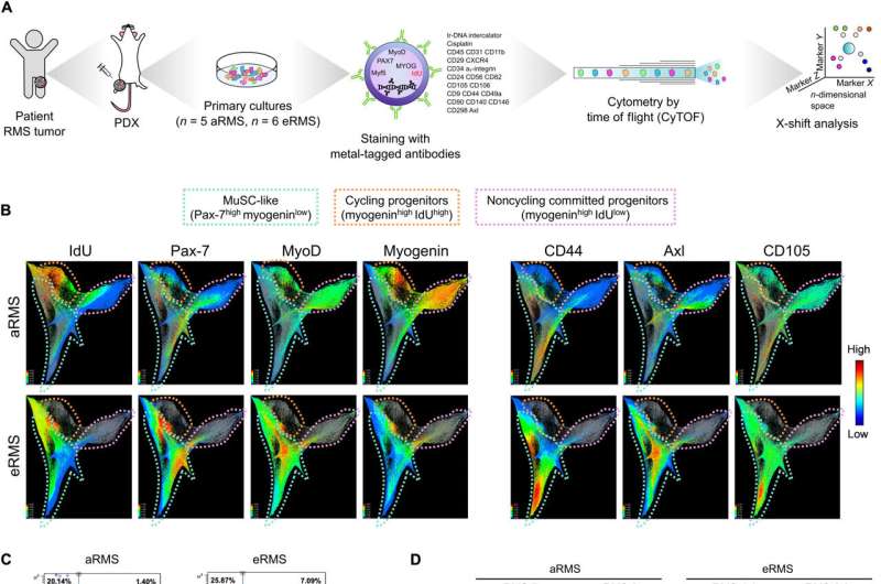
 Experimental workflow. (B) UMAP plot of 48,859 RMS cells after regressing the number of count RNA, the percentage of mitochondrial genes, and the run batch effect. Cells are color-coded based on the corresponding sample of origin. (C) UMAP plot of 48,859 RMS cells after integration. Populations identified by Louvain clustering are shown. (D) Dot plot showing expression of lineage-specific marker genes across the different Louvain clusters in aRMS and eRMS samples. (E) Model of skeletal myogenesis with the populations identified in RMS. UMAP plots are colored on the basis of the expression of markers delineating a myogenic lineage progression. (F) UMAP plot of RMS cells after integration and color-coded based on the sample of origin. (G) Relative proportion of Louvain clusters. Data are represented as means ± SEM; ordinary two-way analysis of variance (ANOVA) with uncorrected Fisher's least significant difference (LSD). *P Science Advances (2023). DOI: 10.1126/sciadv.ade9238")